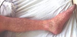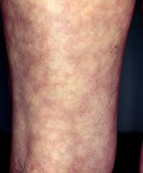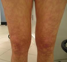

 Livedo reticularis is a mottling of the skin.
Livedo reticularis is associated with autoimmune diseases, hyperlipidemia, poisons, and drug abuse.
Livedo reticularis is a common skin finding consisting of a mottled reticulated vascular pattern that appears as a lace-like purplish discoloration of the skin.
The skin discoloration is caused by ischemia in the arterioles that supply the cutaneous capillaries, resulting in deoxygenated blood showing as the blue discoloration of cyanosis.
This condition increases a person’s risk of forming blood clots including a wide array of pathological and nonpathological conditions.
Disease states include hyperlipidemia, microvascular hematological or anemia states, nutritional deficiencies, hyper- and autoimmune diseases, and drugs/toxins.
Livedo reticularis may be secondary to infrarenal aortoiliac stenosis with severe transient lactic acidosis.
Its differential diagnosis is broadly divided into possible blood diseases, autoimmune, cardiovascular diseases, cancers, and endocrine disorders.
It can be diagnosed in 80% of cases by biopsy.
It may be aggravated by exposure to cold.
It is primarily located in the lower extremities.
Idiopathic livedo reticularis is the most common form of livedo reticularis, completely benign condition of unknown cause affecting mostly young women during the winter.
It is a lacy purple appearance of skin in extremities due to sluggish venous blood flow.
It may be mild, but ulceration may occur.
Secondary livedo reticularis:Reported associated lesions
Vasculitis autoimmune conditions: Livedoid vasculitis Polyarteritis nodosa Systemic lupus erythematosus Dermatomyositis Rheumatoid arthritis Lymphoma Pancreatitis Chronic pancreatitis Tuberculosis Drug-related: Adderall Amantadine Bromocriptine Beta interferon treatment Methylphenidate and dextroamphetamine-induced peripheral vasculopathy Gefitinib Obstruction of capillaries with cryoglobulinaemia Antiphospholipid syndrome due to small blood clots Hypercalcaemia Haematological disorders of polycythemia rubra vera or thrombocytosis Infections-infective endocarditis, syphilis, tuberculosis, Lyme disease Associated with acute kidney injury due to cholesterol emboli status after cardiac catheterization Arteriosclerosis (cholesterol emboli) homocystinuria (due to Chromosome 21 autosomal recessive Cystathionine beta synthase deficiency) Intra-arterial injection Ehlers-Danlos syndrome Pheochromocytoma Livedoid vasculopathy and its association with factor V Leiden mutation FILS syndrome a human syndrome with facial dysmorphism, immunodeficiency, livedo, and short stature. Primary hyperoxaluria, oxalosis Cytomegalovirus infection Generalized livedo reticularis induced by silicone implants for breast augmentation As a rare skin finding in children with Down syndrome Idiopathic livedo reticularis with polyclonal IgM hypergammopathy CO2 angiography Eosinophilic granulomatosis with polyangiiti Erythema nodosum-like cutaneous lesions of sarcoidosis and Sjögren’s syndrome. Livedo vasculopathy associated with IgM antiphosphatidylserine-prothrombin complex antibody Livedo vasculopathy associated with plasminogen activator inhibitor-1 promoter homozygosity and prothrombin G20210A heterozygosity. As a first sign of metastatic breast carcinoma. Livedo reticularis associated with renal cell carcinoma Buerger’s disease Rare manifestation of Graves hyperthyroidism Associated with pernicious anaemia Moyamoya disease Familial primary cryofibrinogenemia.
Livedo reticularis is diagnosed by its clinical appearance and history.
There are no further tests or examinations to confirm the diagnosis of livedo reticularis.
Further investigations may be undertaken where an underlying cause is suspected such as skin biopsies, or blood tests for antibodies associated with antiphospholipid syndrome or systemic lupus erythematosus.
Treatment identifying and treating any underlying conditions in secondary livedo.
First-line treatment typically involves anticoagulants.
Anticoagulants, such as low molecular weight heparin and rivaroxaban, are effective in managing livedo reticularis.
Second-line treatments may include intravenous immunoglobulins (IVIg), especially in cases refractory to anticoagulants.
Additional therapies include antiplatelet agents, anabolic steroids, and hyperbaric oxygen therapy.
Antiplatelet agents like aspirin and pentoxifylline have been used with some success.
Hyperbaric oxygen therapy has been reported to be effective in resistant cases, providing rapid ulcer healing and pain relief.
