 Id reactions are generalized types of acute dermatitis developing after days or weeks at skin locations distant from the initial inflammatory or infectious site.
They can be localized or generalized.
It is also known as an autoeczematous response.
There must be an identifiable initial inflammatory or infectious skin problem which leads to the generalised eczema.
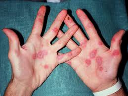
Lesions are intensely itchy, the red papules and pustules can be associated with blisters and scales and are always remote from the primary lesion.
Id reaction is is most commonly a blistering, itchy vesicular rash on the sides of fingers and feet as a reaction to fungal infection on the feet, athlete’s foot.
Possible triggers of the id reaction include: Stasis dermatitis, allergic contact dermatitis, acute irritant contact eczema and infective dermatitis
Id reactions also exist as erythema nodosum, erythema multiforme, erythema multiforme, Sweet’s syndrome, and urticaria.
Id syndrome can be caused by infection with dermatophytosis, mycbacterium, viruses, bacteria and parasites.
Eczema,contact allergic dermatitis, stasis dermatitis, stitches and trauma have been associated with id reactions.
Radiation treatment of tinea capitis can rigger an id reaction.
Potential explanations of pathogenesis include:
Atypical immune recognition of autologous skin antigens
Stimulation of T cells by changing skin
Lower threshold for skin irritation
Spreading of infectious antigens causing a secondary response
Hematogenous dissemination of cytokines from the primary site of inflammation
The id reaction often presents with symmetrical red patches of eczema with papules and vesicles.
Lesions are particularly located on the outer sides of the arms, face and trunk.
They occur suddenly and are intensely itchy, occuring a few days to a week after the initial allergic or irritant dermatitis.
The initial skin problem and the id reaction must be observed to make the diagnosis.
A skin biopsy is rarely necessary for diagnosis.
Biopsy, if done, mostly shows an interstitial granulomatous dermatitis, some lesions being spongiotic, and cannot be distinguished from other skin diseases by histopathology.
Differential diagnosis: include atopic dermatitis, contact dermatitis, dyshidrosis, photodermatitis, scabies and drug eruptions.
Treatment
Id reactions are frequently unresponsive to corticosteroid therapy, but clear when the focus of infection or infestation is treated.
Sometimes medications are used to relieve symptoms, include topical corticosteroids, and antihistamines.
If opportunistic bacterial infection occurs, antibiotics may be required.
A full recovery is expected with treatment,but recurrent id reactions are frequently due to inadequate treatment of the primary infection or dermatitis and often the cause of recurrence is unknown.
Has no particular affinity to any particular ethnic group, seen in all age groups and equally among males and females,.
Its precise prevalence is not known.