1872
2098
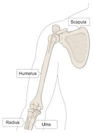
The humerus is the long bone in the upper arm or forelimb that runs from the shoulder to the elbow.
It is the third largest long bone in the human body and plays a crucial role in the function of the upper limb, including movement and support.
The head of the humerus articulates with the glenoid cavity of the scapula to form the shoulder joint.
The proximal end also includes the greater and lesser tubercles, which serve as attachment sites for shoulder muscles.
The humerus articulates proximally with the scapula at the glenohumeral joint, shoulder joint, and distally with the radius and ulna at the elbow joint.
Structurally, it consists of a proximal end, including the head, anatomical neck, and greater and lesser tubercles, a shaft (diaphysis), and a distal end, including the medial and lateral epicondyles, trochlea, and capitulum.
The numeral shaft is the long, cylindrical part of the bone extends down the arm.
The distal end of the humerus articulates with the radius and ulna bones of the forearm at the elbow joint.
Key features include the medial and lateral epicondyles and the trochlea and capitulum, which facilitate joint movement.
The humerus connects the scapula and the two bones of the lower arm, the radius and ulna, and consists of three sections.
The head of the humerus is a rounded structure that articulates with the glenoid cavity of the scapula to form the shoulder joint.
Its anatomical neck, separating it from the tubercles.
The greater and lesser tubercles: Serve as attachment points for rotator cuff muscles.
The intertubercular groove (bicipital groove) lies between them.
The surgical neck is located below the tubercles; a common fracture site.
Humeral shaft
Shape: Cylindrical proximally, becoming flattened distally.
Deltoid Tuberosity: A lateral prominence for deltoid muscle attachment.
Radial Groove: Houses the radial nerve and profunda brachii artery on the posterior side.
Distal humerus: Epicondyles: Medial and lateral projections for muscle attachment.
Condyle: Includes:
Trochlea: Articulates with the ulna. Capitulum: Articulates with the radius. Fossae (coronoid, radial, olecranon) accommodate joint movements during elbow flexion and extension.
The humerus plays a critical role in arm movement and muscle attachment.
The humeral upper extremity consists of a rounded head, a narrow neck, and two short processes, tubercles.
The body of the humerus is cylindrical in its upper portion, and more prismatic below.
The lower extremity bone consists of 2 epicondyles, 2 processes (trochlea and capitulum), and 3 fossae (radial fossa, coronoid fossa, and olecranon fossa).
Its anatomical neck, the constriction below the greater and lesser tubercles of the humerus is referred to as its surgical neck due to its tendency to fracture.
The upper/proximal extremity of the humerus consists of the bone’s large rounded head joined to the body by a constricted portion called the neck, and two eminences, the greater and lesser tubercles.
The head (caput humeri), is nearly hemispherical in form.
It is directed upward, medialward, and a little backward, and articulates with the glenoid cavity of the scapula to form the glenohumeral joint or shoulder joint.
The circumference of its articular surface is slightly constricted and is termed the anatomical neck, in contradistinction to a constriction below the tubercles called the surgical neck which is frequently the seat of fracture.
Fracture of the anatomical neck rarely occurs.
The diameter of the humeral head is generally larger in men than in women.
The anatomical neck of the humerus is obliquely directed, forming an obtuse angle with the body.
In the upper half it is represented by a narrow groove separating the head from the tubercles.
The line separating the head from the rest of the upper end is called the anatomical neck.
The anatomical neck affords attachment to the articular capsule of the shoulder-joint, and is perforated by numerous vascular foramens.
Fracture of the anatomical neck rarely occurs.
The anatomical neck of the humerus is an indentation distal to the head of the humerus on which the articular capsule attaches.
The surgical neck is a narrow area distal to the tubercles that is a common site of fracture.
It makes contact with the axillary nerve and the posterior humeral circumflex artery.
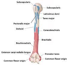
The greater tubercle is a large, posteriorly placed projection that is placed laterally. The greater tubercle is where supraspinatus, infraspinatus and teres minor muscles are attached.
The crest of the greater tubercle forms the lateral lip of the bicipital groove and is the site for insertion of pectoralis major.
The greater tubercle is just lateral to the anatomical neck.
Its upper surface is rounded and marked by three flat impressions.
The impressions: the highest of these gives insertion to the supraspinatus muscle; the middle to the infraspinatus muscle; the lowest one, is the teres minor muscle.
The lesser tubercle is smaller, and anterolaterally placed to the head of the humerus.
The lesser tubercle provides insertion to subscapularis muscle.
The crest of the lesser tubercle forms the medial lip of the bicipital groove and is the site for insertion of teres major and latissimus dorsi muscles.
The lesser tuberosity, is more prominent than the greater: it is situated in front, and is directed medialward and forward.
Above and in front it presents an impression for the insertion of the tendon of the subscapularis muscle.
The tubercles are separated from each other by a deep groove, the bicipital groove.
The bicipital groove lodges the long tendon of the biceps brachii muscle and transmits a branch of the anterior humeral circumflex artery to the shoulder-joint.
The upper part of the humerus is covered with a thin layer of cartilage, lined by the synovial membrane of the shoulder-joint.
Its lower portion gives insertion to the tendon of the latissimus dorsi muscle.
The body or shaft of the humerus is triangular to cylindrical and is compressed anteroposteriorly.
The body or shaft has 3 surfaces, namely: Anterolateral surface: the area between the lateral border of the humerus to the line drawn as a continuation of the crest of the greater tubercle.
The antero-lateral surface gives origin to part of the brachialis.
About the middle of this surface is a rectangular elevation, the deltoid tuberosity for the insertion of the deltoid muscle; below this is the radial sulcus, directed obliquely from behind, forward, and downward, and transmitting the radial nerve and profunda artery.
The antero-medial surface gives insertion to the tendon of the latissimus dorsi muscle; its middle part for the attachment of some of the fibers of the tendon of insertion of the coracobrachialis muscle, and the lower part is gives origin to the brachialis muscle.
Posterior surface is nearly covered by the lateral and medial heads of the Triceps brachii, the former arising above, the latter below the radial sulcus.
Its three borders are:
Anterior: the anterior border runs from the front of the greater tubercle above to the coronoid fossa below, separating the antero-medial from the antero-lateral surface.
The crest of the greater tubercle serves for the insertion of the tendon of the pectoralis major muscle.
At its center it forms the anterior boundary of the deltoid tuberosity, on which the deltoid muscle attaches; below, it is smooth and rounded, affording attachment to the brachialis muscle.
The lateral border runs from the back part of the greater tubercle to the lateral epicondyle, and separates the anterolateral from the posterior surface.
Its upper half is rounded and indistinctly marked, serving for the attachment of the lower part of the insertion of the teres minor muscle, and below this giving origin to the lateral head of the triceps brachii muscle; its center is traversed by a broad but shallow oblique depression, the spiral groove.
The radial nerve runs in the spiral groove.
The lateral border lower part forms a prominent, margin, which presents an anterior lip for the origin of the brachioradialis muscle two-thirds above, and extensor carpi radialis longus muscle one-third below, a posterior lip for the triceps brachii muscle, and an intermediate ridge for the attachment of the lateral intermuscular septum.
The medial border extends from the lesser tubercle to the medial epicondyle.
Its upper third consists of a prominent ridge, the crest of the lesser tubercle, which gives insertion to the tendon of the teres major muscle.
About the humerus center is a slight impression for the insertion of the coracobrachialis muscle, and just below this is the entrance of the nutrient canal, directed downward; sometimes there is a second nutrient canal at the commencement of the radial sulcus.
The inferior third of this medial border is raised into a slight ridge, the medial supracondylar ridge, which became very prominent below, as an anterior lip for the origins of the brachialis muscle and the pronator teres muscle, a posterior lip for the medial head of the triceps brachii muscle, and an intermediate ridge for the attachment of the medial intermuscular septum.
The deltoid tuberosity is a roughened surface on the lateral surface of the shaft of the humerus and acts as the site of insertion of deltoideus muscle.
At the posterorsuperior part of the shaft crest is where the lateral head of triceps brachii is attached.
Deltoid tuberosity of the humerus The radial sulcus, also known as the spiral groove is found on the posterior surface of the shaft and is a shallow oblique groove through which the radial nerve passes along with deep vessels. This is located posteroinferior to the deltoid tuberosity. The inferior boundary of the spiral groove is continuous distally with the lateral border of the shaft.
Radial groove continuing as the lateral border of shaft of the humerus The nutrient foramen of the humerus is located in the anteromedial surface of the humerus. The nutrient arteries enter the humerus through this foramen.
Nutrient foramen of the humerus Distal humerus edit Lower extremity of humerus
Left humerus. Anterior view. Details Identifiers Latin extremitas distalis humeri MeSH D006811 TA98 A02.4.04.001 TA2 1180 FMA 13303 Anatomical terms of bone [edit on Wikidata] The distal or lower extremity of the humerus is flattened from before backward, and curved slightly forward; it ends below in a broad, articular surface, which is divided into two parts by a slight ridge. Projecting on either side are the lateral and medial epicondyles. Articular surface edit The articular surface extends a little lower than the epicondyles, and is curved slightly forward; its medial extremity occupies a lower level than the lateral. The lateral portion of this surface consists of a smooth, rounded eminence, named the capitulum of the humerus; it articulates with the cup-shaped depression on the head of the radius, and is limited to the front and lower part of the bone. Fossae edit Above the front part of the trochlea is a small depression, the coronoid fossa, which receives the coronoid process of the ulna during flexion of the forearm. Above the back part of the trochlea is a deep triangular depression, the olecranon fossa, in which the summit of the olecranon is received in extension of the forearm.
Olecranon fossa of the humerus The coronoid fossa is the medial hollow part on the anterior surface of the distal humerus. The coronoid fossa is smaller than the olecranon fossa and receives the coronoid process of the ulna during maximum flexion of the elbow.
Coronoid fossa of the humerus Above the front part of the capitulum is a slight depression, the radial fossa, which receives the anterior border of the head of the radius, when the forearm is flexed.
Radial fossa of the humerus These fossæ are separated from one another by a thin, transparent lamina of bone, which is sometimes perforated by a supratrochlear foramen; they are lined in the fresh state by the synovial membrane of the elbow-joint, and their margins afford attachment to the anterior and posterior ligaments of this articulation. The capitulum is a rounded eminence forming the lateral part of the distal humerus. The head of the radius articulates with the capitulum.
Capitulum on the lateral side and trochlea on the medial side of the humerus The trochlea is spool-shaped medial portion of the distal humerus and articulates with the ulna.
The epicondyles are continuous with the supracondylar ridges.
The lateral epicondyle is a small, tuberculated eminence, curved and gives attachment to the radial collateral ligament of the elbow-joint, and to a tendon common to the origin of the supinator and some of the extensor muscles.
The medial epicondyle, gives attachment to the ulnar collateral ligament of the elbow-joint, to the pronator teres, and to a common tendon of origin of some of the flexor muscles of the forearm; the ulnar nerve runs in a groove on the back of this epicondyle.
At the shoulder, the head of the humerus articulates with the glenoid fossa of the scapula.
More distally, at the elbow, the capitulum of the humerus articulates with the head of the radius, and the trochlea of the humerus articulates with the trochlear notch of the ulna.
The axillary nerve is located at the proximal end, against the shoulder girdle.
Dislocation of the humerus’s glenohumeral joint has the potential to injure the axillary nerve or the axillary artery.
Signs and symptoms of this dislocation include a loss of the normal shoulder contour and a palpable depression under the acromion.
The radial nerve follows the humerus closely.
At the midshaft of the humerus, the radial nerve travels from the posterior to the anterior aspect of the bone in the spiral groove.
A fracture of the humerus in this region can result in radial nerve injury.
The ulnar nerve lies at the distal end of the humerus near the elbow.
When struck, it can cause a distinct tingling sensation, and sometimes a significant amount of pain.
It lies posterior to the medial epicondyle, and is easily damaged in elbow injuries.
The deltoid muscle originates on the lateral third of the clavicle, acromion and the crest of the spine of the scapula.
It is inserted on the deltoid tuberosity of the humerus and has several actions including abduction, extension, and circumduction of the shoulder.
The supraspinatus also originates on the spine of the scapula. It inserts on the greater tubercle of the humerus, and assists in abduction of the shoulder.
The pectoralis major, teres major, and latissimus dorsi insert at the intertubercular groove of the humerus.
They work to adduct and medially, or internally, rotate the humerus.
The infraspinatus and teres minor insert on the greater tubercle, and work to laterally, or externally, rotate the humerus.
The subscapularis muscle inserts onto the lesser tubercle and works to medially, or internally, rotate the humerus.
The biceps brachii, brachialis, and brachioradialis act to flex the elbow.
The biceps does not attach to the humerus.
The triceps brachii and anconeus extend the elbow, and attach to the posterior side of the humerus.
The four muscles of supraspinatus, infraspinatus, teres minor and subscapularis form a musculo-ligamentous girdle called the rotator cuff.
The rotator cuff stabilizes the very mobile but inherently unstable glenohumeral joint.
The other muscles are used as counterbalances for the actions of lifting/pulling and pressing/pushing.
The humerus allows a wide range of arm movements, working in conjunction with muscles, tendons, and ligaments to enable flexion, extension, rotation, and other movements of the arm.
The humerus provides structural support to the arm and serves as an anchor point for important muscles.
Humerus fractures can occur due to trauma, such as falls or direct impacts.
Humerus fractures can be categorized into proximal humerus fractures, mid-shaft fractures, and distal humerus fractures.
Humerus dislocations typically involve the shoulder joint and can occur from sports injuries or accidents.
Common injuries associated with the humerus include fractures of the proximal humerus, humeral shaft, and distal humerus.
Proximal humerus fractures are common and often result from falls in older adults or high-energy trauma in younger individuals.
Humeral fractures can involve the humeral head, greater tuberosity, and lesser tuberosity.
Complications can include rotator cuff tears and avascular necrosis.
Humeral shaft fractures are also common and can result from direct trauma or falls.
These fractures are often managed nonoperatively, but surgical intervention may be required in cases of open fractures, multiple trauma, or significant nerve injuries.
Primary radial nerve palsy is a notable complication, occurring in approximately 10% of cases.
Distal humerus fractures often involve the supracondylar region and are less common but can be more complex, particularly in pediatric populations.
These fractures often can be associated with significant neurovascular injuries.
In children, these fractures are frequently related to sports activities or falls and may require surgical intervention for proper alignment and healing.
Overall, the management of humeral fractures depends on the fracture location, patient age, and associated injuries, with a combination of nonoperative and operative strategies employed based on specific clinical scenarios.
Proximal humerus fractures can be managed nonoperatively or operatively, depending on the fracture type and patient factors.
Nonoperative management includes immobilization and early physical therapy.
Surgical options include open reduction and internal fixation (ORIF) with locking plates, intramedullary nailing, and arthroplasty (hemiarthroplasty or reverse shoulder arthroplasty).
Reverse shoulder arthroplasty is often preferred for complex fractures in older adults due to better functional outcomes.
Humeral shaft fractures are typically treated nonoperatively with functional bracing, achieving high union rates.
Surgical intervention is indicated for open fractures, polytrauma, or failure of nonoperative management.
Surgical options include ORIF with plates, intramedullary nailing, and external fixation.
Plate fixation is associated with higher union rates and better functional outcomes compared to intramedullary nailing, though it carries a higher risk of radial nerve palsy.
Distal humerus fractures often require surgical management due to their complexity.
ORIF with dual plating is the gold standard, providing stable fixation and allowing early mobilization.
Nonoperative management is reserved for non-displaced fractures or patients with significant comorbidities.