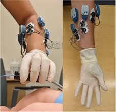
 Electromyoneurography (EMNG) refers to the combined use of electromyography and electroneurography.
It measures a peripheral nerve’s conduction velocity upon stimulation alongside electrical recording of muscular activity.
Electromyogram (EMG) measures the electrical activity in the muscle. EMG can show nonspecific signs of muscle damage or irritability.
Nerve conduction velocity (NCV) measures the how fast signals travel along a nerve.
The nerve signals are measured with surface electrodes or needle electrodes.
It allows for both the source and location and for more accurate diagnosis of a particular neuromuscular disease.
Electromyoneurography is a technique that uses surface electrical probes to obtain electrophysiological readings from nerve and muscle cells.
Nerve activity is recorded using surface electrodes, stimulating the nerve at one site and recording from another with a minimum distance between the two: time difference of the potential is a measure of the time taken for the potential to travel the distance across the two sites.
This measures the conduction velocity along the nerve.
The amplitude of the potential, measures the number of fibers conducting the response.
Absence or low amplitude, indicates potential nerve damage.
It is used is to detect neuropathy due to diseases like diabetes mellitus, muscle weakness or paralysis due to sepsis or multi-organ failure in comatose patients, and neuromuscular abnormalities.
The procedure analyzes the nerve conduction and muscle potentials through the use of H-Reflex and F-Wave studies.
Findings are measured in the form of amplitude (mV), latency (ms), and velocity (m/s) of the injured radial nerve, before and after surgery.
It has a high level of sensitivity for detecting peripheral nerve damage and myopathies.
It has diagnostic capabilities when diagnosing peripheral neuropathy disorders like radiculopathy, and axonopathy in addition to myopathies such as muscular dystrophy, myotonia, and myasthenia gravis.
It is used to detect diabetic polyneuropathy, a serious condition that is progressive in nature.
It can be used to measure recovery from a surgical procedure on a nerve.
Electromyoneurography’s results in a higher level of diagnostic ability and often results in a lesser demand for more invasive techniques for acquiring electrophysiological data, such as myelography.
Electromyoneurography useful in diagnosing the following neuromuscular conditions:
Myopathy
Neuropathy
Primary muscular dystrophy
Duchenne muscular dystrophy
Facioscapulohumeral muscular dystrophy
Limb-girdle muscular dystrophy
Myelopathy-a lesion involving motor neuron in anterior horn of the spinal cord.
Spinal muscular atrophy
Progressive muscular atrophy
Poliomyelitis
Amyotrophic lateral sclerosis
Charcot–Marie–Tooth disease
Cell membrane hyper-irritability
Myotonic dystrophy
Myotonia congenita
Paramyotonia congenita
Radiculopathy-lesions involving the nerve root.
Spinal disc herniation
Spinal stenosis
Guillain–Barré syndrome
Myasthenia
Myasthenia gravis
Lambert–Eaton myasthenic syndrome
Hypokalemia
Glycogen storage disease type V
Cushing’s syndrome
Axonopathy- disease or damage to the axon or peripheral nerve
Carpal tunnel syndrome
Radial neuropathy
Meralgia paraesthetica
Hypothyroidism
In EMNG muscle recordings is done by insertion of a needle, when the muscle is at rest and when the muscle is contracting.
In addition to examining the muscles, the conduction velocity of nerve signals are measured.
The nerve’s ability to transmit signals is tested
Needles inserting recording electrodes capture the data and signal electrodes to initiate signals down a nerve by applying a small shock.
Evaluating a nerve’s conduction velocity, and testing potentials, allows for a diagnosis that can detect pain and sensory problems at the neuromuscular level.
The needle is attached to a recording device, an electromyography machine indicating action potential or graded potential spikes.
Interpretation indicates a decreased amplitude or duration may indicate nerve damage due to a muscle diseases, whereas an increase in these demonstrates reinervation, or repair by new nerve connections to the muscles, has occurred.
