 The cingulate cortex is a part of the brain situated in the medial aspect of the cerebral cortex.
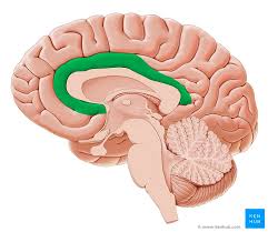
The cingulate cortex includes the entire cingulate gyrus, which lies immediately above the corpus callosum, and the continuation of this in the cingulate sulcus.
The cingulate cortex is usually considered part of the limbic lobe.
The cingulate cortex receives inputs from the thalamus and the neocortex, and projects to the entorhinal cortex via the cingulum.
It is an integral part of the limbic system, which is involved with emotion formation and processing, learning, and memory.
The cingulate gyrus highly influential in linking motivational outcomes to behavior induces a positive emotional response, which results in learning.
The cingulate cortex highly important in disorders such as depression and schizophrenia, and plays a role in executive function and respiratory control.
Based on cerebral cytoarchitectonics it has been divided into the Brodmann areas 23, 24, 26, 29, 30, 31, 32 and 33.
The areas 26, 29 and 30 are usually referred to as the retrosplenial areas.
The cingulate cortex plays important roles in various cognitive and emotional functions.
It’s part of the limbic system and forms a collar-like region around the corpus callosum, which connects the brain’s hemispheres.
The cingulate cortex is typically divided into several regions:
Anterior cingulate cortex (ACC) – Involved in attention allocation, reward anticipation, decision-making, ethics, impulse control, and emotion regulation.
Midcingulate cortex – Often associated with response selection and motor control.
Posterior cingulate cortex (PCC) – Linked to episodic memory, visuospatial processing, and consciousness.
Functionally, the cingulate cortex serves as an integration hub that connects cognitive, attentional, and emotional processes. It’s particularly important for:
– Emotional processing and regulation – Pain processing – Learning and memory – Cognitive control and executive function – Self-reference and introspection
Dysfunction in the cingulate cortex has been implicated in various disorders including depression, anxiety disorders, obsessive-compulsive disorder, and attention deficit hyperactivity disorder.
The ACC is involved in error and conflict detection processes.
Outputs of the posterior cingulate gyrus
The posterior cingulate cortex (Brodmann’s area 23) sends projections to dorsolateral prefrontal cortex (Brodmann’s area 9), anterior prefrontal cortex (Brodmann’s area 10), orbitofrontal cortex (Brodmanns’ area 11), the parahippocampal gyrus, posterior part of the inferior parietal lobule, the presubiculum, the superior temporal sulcus and the retrosplenial region.
The rostral anterior cingulate gyrus is larger in healthy females than males, but this sex difference was not found in people with schizophrenia.
The metabolic rate of glucose was found to be lower in the left anterior cingulate gyrus and the right posterior cingulate gyrus in people with schizophrenia relative to controls.
The volume of the left anterior cingulate gyrus was reduced in people with schizophrenia.
The smaller the size of anterior cingulate gyrus, the lower was the level of social functioning and the higher was the psychopathology in the people with schizophrenia.
The anterior cingulate gyrus was found to be bilaterally smaller in people with schizophrenia as compared with control group.
No difference in IQ tests and basic visuoperceptual ability with facial stimuli was found between people with schizophrenia and the control.
The cingulate gyrus is heavily implicated in depressive disorders.
Isolated stroke of the cingulate gyrus has also been found to induce depression, potentially implicating this region in post-stroke depression which may occur following stroke of a larger part of the brain.
Greater connectivity in the cingulo-opercular network is linked to better episodic memory, attention, and executive function, but declines with age, impacting cognitive performance: Reduced connectivity in the anterior cingulate cortex within the salience network correlates with cognitive decline.
Age-related decreases in cingulo-opercular connectivity, especially in the left insula, mediate declines in visual processing speed.