 Bullosis diabeticorum, also known as diabetic bullae or bullous eruption of diabetes mellitus, is a specific type of skin lesion occurring in patients with diabetes mellitus.
Recurrent spontaneous blisters occurring on the extremities of patients with diabetes mellitus.
Rare, occurs in 0.5% of diabetics.
Most habe a long-standing disease with peripheral neuropathy.
The lesions are asymptomatic and healed slowly without scarring in 2 to 6 weeks.
Patients with insulin-dependent diabetes have a marked reduced threshold to suction blister development compared to age-matched normal controls.
There is an acral prominence of BD, so the role of trauma has been speculated.
But trauma does not this cannot for the absence of BD in the vast majority of the diabetic population, with the absence in most cases of any history of trauma and the fact that these lesions resolve spontaneously.
The majority of patients with diabetes mellitus with BD have associated nephropathy and neuropathy, suggesting a microangiopathy and aging of connective tissue and, more precisely, a local subbasement membrane zone connective-tissue alteration.
Postulated disturbances in calcium, magnesium, and carbohydrate metabolism ,and excessive exposure to ultraviolet light, especially in those with nephropathy, have also been suggested as a causative factor.
In a retrospective study of 5000 patients with diabetes mellitus, the incidence of BD was 0.16% per year.
BD more frequently presented in adult men who had long-standing uncontrolled diabetes.
The reported male-to-female ratio is 2:1.
The histologic finding show bullae with inconsistent levels of skin layer separation.
Bullae are initially subepidermal, and then intraepidermal blisters represent older lesions undergoing re-epithelialization.
The adjacent epidermis is often unremarkable.
The dermis shows minimal inflammation, and microvascular changes consistent with diabetes are also present. The
Proteinaceous fluid contained in bullae is, most of the time, clear and sterile.
Direct immunofluorescence examination is usually negative.
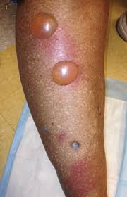
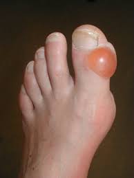
It starts with a sudden appearance of one or more painless, tense vesicles or bullae within normal-appearing, non-inflamed skin, generally on the acral portions of the body.
Most frequent locations are the feet, distal legs, hands, and forearms.
Blisters particularly appear on the tips of the toes and the plantar surface of the feet, but rarely present on the trunk.
The lesions are usually asymptomatic, but occasionally notice a mild burning sensation.
Blisters often develop in the absence of known trauma.
The vesicles and bullae range in size from few millimeters to several centimeters.
Lesions typically start as tense blisters, that contain a clear and sterile fluid, which may sometimes be hemorrhagic.
Most patients with BD have longstanding diabetes.
It is associated with both insulin-dependent and noninsulin-dependent diabetes mellitus, and patients have an associated polyneuropathy, retinopathy, or nephropathy.
Occcasionally bullae can be a presenting sign of the disease.
Management:
The blisters are usually self-limiting, with bullae said to resolve untreated within 2 to 6 weeks.
Some advise that the blistered skin should be left intact since it constitutes an effective and sterile cover for the underlying wound.
Some advocate a small-bore needle aspiration or placement of a small window in the blister roof and application of topical antiseptics or antibiotics to reduce discomfort and prevent secondary infections.
In some cases, surgical management of BD is required to reduce risk of osteomyelitis.
Differential diagnosis of BD:
porphyria cutanea tarda and pseudoporphyria, the blisters are usually infracentimetric and favor the hands rather than the feet and ankles.
The distal extremity is also a common site for erythema multiforme and fixed drug eruptions.
Epidermolysis bullosa acquisita and localized bullous pemphigoid, closely resembling BD.
BD is more frequently a self-limiting condition and usually resolves spontaneously within a few weeks.
Lesions typically heal without post-inflammatory pigmentation or residual scarring.
However, repeated and recurrent episodes can lead to ulceration, and scarring.
Bullous symptoms can reoccur over the years.
Treatment remains mainly supportive.