
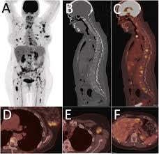
 FDG-PET/CT in breast cancer is primarily a staging tool for higher-risk, locally advanced, or recurrent disease, and not a routine test for early-stage cancer.
Its greatest value is detecting extra-axillary regional nodal disease and unsuspected distant metastases, which can shift management from curative-intent combined-modality therapy to systemic/palliative treatment. It has essentially no added value in clinical stage I (T1N0) disease and offers uncertain benefit in stage IIA.[1][2]
NCCN indications (v6.2026). FDG-PET/CT is listed as an optional additional test “as clinically indicated” during workup prior to preoperative systemic therapy for c≥T2 or cN+ (M0) disease, cT1c/cN0 HER2-positive disease, or cT1c/cN0 triple-negative disease.[3] Key points from the algorithm and its footnotes:
– It is most beneficial and accurate for stage III and invasive ductal (vs. lobular) histology, but may be useful in selected stage IIA cases (cT1cN1, cT2cN0) — e.g., equivocal CT/bone scan, suspicion of undetected nodal/distant disease, or treatment-response assessment.[3]
– It may be used as an adjunct to, or in lieu of, standard staging and can be performed simultaneously with diagnostic CT.[3]
– If upfront FDG-PET/CT clearly shows concordant PET and CT findings, a bone scan or sodium fluoride PET/CT may not be needed.[3]
– For inflammatory breast cancer, FDG-PET/CT is offered as an alternative to bone scan in the workup, and is particularly helpful when standard staging is equivocal or for identifying unsuspected regional nodal/distant disease.[3]
Here is the NCCN workup algorithm showing where FDG-PET/CT fits in the pre-treatment evaluation:
FDG-PET/CT has a defined role in baseline staging from stage IIB through stage IV, is not recommended in stage I, and is only possibly useful in stage IIA without enough data for routine use.
In newly diagnosed stage IIB–III disease, PET/CT modifies stage in roughly 14–28% of patients regardless of receptor status, with sensitivity/specificity for distant metastases of ~97%/91% versus ~86%/67% for conventional imaging.
Upstaging yield rises steeply with baseline stage: unsuspected metastases were found in ~2% of stage IIA, 11% of IIB, 18% of IIIA, 37% of IIIB, and 47% of IIIC in one series.
A meta-analysis of 29 studies (4,276 patients) found stage change in 25% and management change in 18%, increasing from 11% (stage I) to 20% (II) to 34% (III).
The randomized PETABC trial (Dayes et al., stage IIb T3N0 or III) is the strongest evidence: PET/CT upstaged 23% to stage IV versus 11% with conventional staging (bone scan + CT), an absolute difference of 12.3%, and fewer PET/CT patients received combined-modality therapy (81% vs 89%).
Recurrence/restaging: More effective than conventional imaging at detecting locoregional or distant recurrence, including when tumor markers (CA 15-3, CEA) are elevated but conventional imaging is equivocal, and useful for whole-body workup to determine whether recurrence is isolated.
Bone metastases: More sensitive/specific than bone scan for lytic/mixed lesions and marrow involvement; FDG uptake is more variable in osteoblastic lesions, where the CT component aids detection.
May assess early metabolic response, particularly in non-metastatic TNBC and HER2-positive tumors, using PERCIST/EORTC criteria on certified scanners.
Limitations. Sensitivity is lower for invasive lobular carcinoma and for low-grade, low-proliferative, well-differentiated luminal tumors.
PET/CT cannot replace surgical axillary/sentinel node staging in early disease — a negative axilla on PET does not exclude microscopic nodal involvement (it is a rule-in, not rule-out, tool for nodal disease).
PET is also less sensitive than dedicated chest CT for small lung nodules because of partial-volume and respiratory-motion effects.
Repeated PET scan after two weeks of treatment may improve outcome estimation in newly diagnosed metastatic breast cancer patients compared to standard CT alone: patients without disease progression on early PET scan had a better median progression free survival and overall survival than patients with disease progression: After eight weeks those with progression on early PET scan had a median overall survival of 22.3 months, whereas patients without progression on PET scan had overall survival of 50.1 months (IMPACT Metastatic Breast Cancer consortium).