 Amyloid microclots (or fibrinaloid microclots) are small, abnormal clumps of misfolded proteins—primarily fibrinogen and fibrin—that form in the bloodstream.
Amyloid microclots are anomalous circulating aggregates in which fibrinogen polymerizes into a β-sheet-rich, amyloid-like form of fibrin that is resistant to normal fibrinolysis.
They are distinct from canonical thrombi: rather than a typical crosslinked fibrin-platelet mesh, they are misfolded protein aggregates that entrap inflammatory molecules, antibodies, and other plasma proteins.
They are highly resistant to the body’s natural ability to break down clots and are a major area of research in conditions involving chronic inflammation.
There body’s clotting system activates proteins like fibrinogen to form temporary blood clots.
Normal fibrinogen, when triggered by inflammation or other stimuli (including certain bacterial products, spike proteins, or inflammatory cytokines), can misfold into a beta-sheet-rich amyloid structure.
These anomalous clots are: Extremely dense and compact Resistant to fibrinolysis Able to trap inflammatory molecules, platelets, and other proteins
In the presence of intense systemic inflammation, infections (like SARS-CoV-2), or other triggers, the structure of these proteins can be forced to misfold into an “amyloid” state similar to the protein plaques seen in Alzheimer’s disease.
These misfolded proteins then clump together into microclots that are often wrapped in DNA and inflammatory molecules, making them difficult for the body to dissolve.
Normally, fibrin forms a mesh-like clot during coagulation.
Under certain pathological conditions, fibrinogen can misfold into an amyloid-like structure, creating dense, highly resistant microclots that are difficult for the body to dissolve.
These microclots can range from 1 to 200 microns in size, they can circulate and get trapped in tiny capillaries. restricting blood flow and preventing oxygen from reaching vital tissues.
They obstruct small capillaries and reduce oxygen delivery to tissues
They persist in circulation and cause ongoing inflammation, contribute to platelet hyperactivation, and Impair organ function over time
This systemic microvascular blockage and resulting tissue hypoxia are theoretical primary drivers of several symptoms, including:Debilitating fatigue and brain fog, post-exertional malaise, autonomic nervous system dysfunction, including, postural orthostatic tachycardia syndrome (POTS).
The central hypothesis is that these fibrinolysis-resistant microclots can obstruct microcapillaries, impairing red blood cell transit and oxygen exchange, thereby contributing to symptoms such as fatigue, brain fog, and dyspnea.
Amyloid microclots are being studied in the context of Long COVID (PASC), in Myalgic encephalitis chronic fatigue syndrome (ME/CFS), sepsis, and other chronic inflammatory or metabolic diseases like diabetes and Alzheimer’s.
Detection:
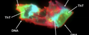
Microclots are typically visualized using fluorescence microscopy with amyloid-sensitive dyes (like thioflavin T) on platelet-poor plasma samples — not standard clinical lab tests.
These structures are identified by staining platelet-poor plasma (PPP) with thioflavin T (ThT), a fluorogenic amyloid dye, and visualizing them under fluorescence microscopy.
Treatments: standard clot-busting drugs often struggle to break down these amyloid-rich clumps, research is focused on alternative approaches.
Investigational treatments:being studied: triple anticoagulant therapy (dual antiplatelet drugs combined with direct oral anticoagulants) and non-invasive methods like low-intensity ultrasound therapy.
Research is ongoing withanticoagulants, anti-inflammatory agents, and fibrinolytic therapies.
Researchers have found abundant amyloid microclots in Long COVID patients’ blood, potentially explaining fatigue, brain fog, and exercise intolerance.
Acute COVID-19 Similar microclots were found in acute infection, correlating with severity and thrombotic complications.
Long COVID patients have abundant anomalous amyloid microclots in their blood, even in the absence of acute infection.
These microclots can trap inflammatory molecules (like cytokines and complement proteins) and are thought to contribute to the persistent symptoms — fatigue, brain fog, and breathlessness — by impairing microcirculation and oxygen delivery.
Myalgic encephalitis/chronic-fatigue syndrome— Similar findings have been reported, suggesting possible overlap.
Amyloid microclots have been observed in diabetic patients.
Cardiovascular disease — Implicated in thromboembolic complications
Alzheimer’s disease — Fibrin amyloid deposits are found in brain vasculature
A Cochrane review noted that the term “microclot” is somewhat of a misnomer, as these are not true thrombi but rather amyloid fibrin(ogen) particles — misfolded, aggregating protein complexes containing cross-β structures.
Proteomic analysis has confirmed they consist of amyloid-fibrinogen aggregates along with entrapped molecules such as α-2-antiplasmin, platelet factor 4 (PF4), von Willebrand factor (VWF), and various antibodies.
Resistance to fibrinolysis: The amyloid conformation renders them resistant to plasmin-mediated degradation. SARS-CoV-2 spike protein amyloid fibrils (particularly the Spike685 peptide, sequence 685–701) have been shown to induce dense fibrin networks resistant to plasmin lysis in vitro.
Proteomic studies show reduced plasma kallikrein and increased PF4 and VWF within microclots, suggesting a self-perpetuating cycle of failed fibrinolysis and platelet hyperactivation.
Structural association with NETs: Circulating microclots are structurally linked to neutrophil extracellular traps (NETs), with Long COVID patients showing elevated microclot counts across all size ranges alongside increased cell-free DNA, myeloperoxidase, and neutrophil elastase.
Procoagulant surface generation: Phosphatidylserine externalization and glycocalyx damage during inflammation create amyloidogenic surfaces that attract coagulation factors and promote prothrombinase assembly, driving formation of fibrin monomer complexes.
Fibrin amyloid microclots and platelet pathology have been identified in blood samples from Long COVID patients, correlating with persistent symptoms including fatigue (74%), cognitive impairment (71%), and dyspnea.
In a study of 104 ICU patients, microclots were detected in 42.3% on admission and were significantly associated with sepsis.
Inflammatory conditions (e.g., rheumatoid arthritis, lupus) are associated with amyloid microclots.
High microclot burden predicted DIC and 28-day mortality.
Type 2 Diabetes Amyloid fibrin clots have been documented in diabetic patients, potentially contributing to the elevated cardiovascular and thrombotic risk seen in this population.
Alzheimer’s disease: Beta-amyloid peptide interacts with fibrinogen to form structurally abnormal, fibrinolysis-resistant clots.
There is evidence that fibrin(ogen) deposits in the brain interact with amyloid-beta (Aβ) plaques, and that this fibrin-amyloid interaction promotes neuroinflammation and vascular damage, potentially worsening disease progression.
Amyloid microclots may:
Obstruct small blood vessels (capillaries) Trap pro-inflammatory proteins Trigger ongoing immune activation Impair tissue oxygenation
Autoimmunity: The anomalous polymerization generates novel epitopes on normal proteins, potentially triggering autoantibody production — a proposed mechanism linking fibrinaloid formation to post-infectious autoimmunity.
Proposed interventions remain largely investigational:
Triple anticoagulant therapy (dual antiplatelet + anticoagulant) has been reported in preliminary, uncontrolled studies to reduce microclot burden and improve Long COVID symptoms.
No validated, guideline-endorsed treatment targeting amyloid microclots currently exists.
Microclots are poorly characterized, detection methods lack standardization, and the causal relationship between microclot presence and clinical symptoms has not been established through rigorous clinical trials.
Standard coagulation and inflammatory markers (CRP, D-dimer) may not reflect the microclot burden, which has contributed to diagnostic uncertainty.
Anomalous clot structure is associated with increased risk of thrombosis and poor outcomes after events like myocardial infarction.
Chronic inflammation can trigger fibrin misfolding, and abnormal clot structure has been observed in these conditions.
The key concern is that amyloid fibroclots are resistant to normal enzymatic breakdown (fibrinolysis), meaning they can persist and accumulate.
Therapeutic Implications
This has led to interest in fibrinolytic therapies (e.g., nattokinase, lumbrokinase, low-dose anticoagulants) as potential treatments, particularly in Long COVID — though clinical evidence is still emerging and these approaches remain experimental.
This is an active and rapidly evolving research area, with the fibrin-amyloid connection now seen as a potential common thread across several chronic inflammatory and thrombotic diseases.