 The epididymis is a long, tightly coiled tube located on the posterior surface of each testicle.
The human epididymis is situated posterior and somewhat lateral to the testis.
It acts as a critical intermediary in the male reproductive system, connecting the testicle to the vas deferens.
It appears as a small, tightly coiled comma-shaped structure about 3.8 to 5 cm long, its internal tubing would measure approximately 6 meters (20 feet) if uncoiled.
The epididymis is invested completely by the tunica vaginalis.
It connects the testicle to the vas deferens in the male reproductive system.
The epididymis is a conduit, and is an active participant in male fertility:
Sperm Maturation: Immature sperm entering from the testes gain motility and fertilize an egg as they transit through the tube.
The distal portion (the tail) serves as the primary reservoir for mature sperm until ejaculation.
Transport: Rhythmic contractions of its smooth muscle walls move sperm toward the vas deferens over a period of 10 to 15 days.
It creates a unique biochemical environment that protects sperm from oxidative stress and the body’s own immune system.
Its primary function is the storage, maturation and transport of sperm cells.
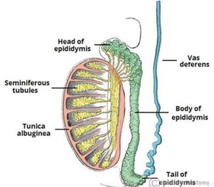
The epididymis is divided into three main regions:
Head (Caput): The widest part at the top of the testicle; it receives sperm from the efferent ducts and begins the process of fluid absorption to concentrate the sperm.
Body (Corpus): The middle section where sperm further mature and begin to acquire motility.
The body has an intermediate epithelium and smooth muscle thickness.
Tail (Cauda): The narrowest part at the bottom, which stores mature sperm and is continuous with the vas deferens.
This has the thinnest epithelium of the three regions and the greatest quantity of smooth muscle.
The tail is distally continuous with the convoluted portion of the ductus deferens/vas deferens.
Symptoms typically include:
Swelling and redness of the scrotum. Gradual onset of testicle pain and tenderness, usually on one side. Painful urination or urgent need to urinate. Blood in the semen.
The epididymis can be divided into three main regions:
The head: The head of the epididymis receives spermatozoa via the efferent ducts of the mediastinum of the testis at the superior pole of the testis.
The head is characterized histologically by a thick epithelium with long stereocilia and a little smooth muscle.
In the caput (head) region these cells have long stereocilia that are tuft-like extensions that project into the lumen.
It is involved in absorbing fluid to make the sperm more concentrated.
Histology The epididymis is covered by a two layered pseudostratified epithelium.
Principal cells: columnar cells that, with the basal cells, form the majority of the epithelium.
The epithelium cells also secrete carnitine, sialic acid, glycoproteins, and glycerylphosphorylcholine into the lumen.
Apical cells: predominantly found in the head region
Clear cells: predominant in the tail region
Intraepithelial lymphocytes: distributed throughout the tissue. Intraepithelial macrophages.
The stereocilia of the epididymis are long cytoplasmic projections that have an actin filament backbone.
The stereocilia in the epididymis are non-motile membrane extensions that increase the surface area of the cell, allowing for greater absorption and secretion.
It has been shown that epithelial sodium channel ENaC that allows the flow of Na+ ions into the cell is localized on stereocilia.
Because sperm are initially non-motile as they leave the seminiferous tubules, large volumes of fluid are secreted to propel them to the epididymis.
The core function of the stereocilia is to resorb 90% of this fluid as the spermatozoa start to become motile.
This absorption creates a fluid current that moves the immobile sperm from the seminiferous tubules to the epididymis.
Spermatozoa only reach full motility when inside a vagina, where the alkaline pH is neutralized by acidic vaginal fluids.
Spermatozoa formed in the testis enter the caput epididymidis, progress to the corpus, and finally reach the cauda region, where they are stored.
Sperm entering the caput epididymidis are incomplete—they lack the ability to swim forward (motility) and to fertilize an egg. Epididymal transit takes 2 to 6 days in humans and 10–13 in rodents.
During their transit in the epididymis, sperm undergo maturation processes necessary for them to acquire motility and fertility.
Final maturation (capacitation) is completed in the female reproductive tract.
The epididymis secretes immobilin, a large glycoprotein that is responsible for the creating of the viscoelastic luminal environment that serves to mechanically immobilize spermatozoa until ejaculation.
Immobilin is predominantly secreted into the proximal caput epididymidis prior to the acquisition of the potential for sperm motility.
During emission, sperm flow from the cauda epididymis, which functions as a storage reservoir, into the vas deferens where they are propelled by the peristaltic action of muscle layers in the wall of the vas deferens, and are mixed with the diluting fluids of the prostate, seminal vesicles, and other accessory glands prior to ejaculation forming semen.
During their transit through the epididymis, the spermatozoa undergo a series of transformations in preparation for their ultimate task of fertilizing the oocyte.
In order to protect the spermatozoa in the epididymis, the epididymal epithelium produces a variety of antioxidant proteins that help protect the spermatozoa from oxidative damage.
The antioxidant proteins produced in the epididymis include catalase, glutathione peroxidases, glutathione-S-transferases, peroxiredoxins, superoxide dismutases, thioredoxin reductase and thioredoxins.
Deficiencies in these antioxidant proteins reduces sperm quality by affecting a variety of the proteins necessary for the motility needed to fertilize oocytes.
Reduced antioxidant activity also causes increased oxidative damage to the sperm DNA.
Epididymitis an inflammation of the epididymis and is much more common than testicular inflammation, termed orchitis.
The most frequent condition affecting this organ is epididymitis, which is inflammation often caused by a bacterial infection or a sexually transmitted infection (STI) like chlamydia or gonorrhea.
Epididymotomy is the placing of an incision into the epididymis and is sometimes used as a treatment option for acute suppurating epididymitis.
Epididymectomy is the surgical removal of the epididymis sometimes performed for post-vasectomy pain syndrome and for refractory cases of epididymitis.