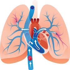
Pulmonary embolism (PE) is defined as an occlusion of the pulmonary arterial tree that impedes blood flow distal to the blockage.
Most cases result from thromboemboli originating in the deep veins of the lower extremities or pelvis.
PE occurs in approximately 1 per 1,000 persons annually worldwide.
In the United States, an estimated 60,000–100,000 deaths occur annually, accounting for 5–10% of all hospital deaths.
25% of patients present with sudden death as the initial manifestation.
The three-month mortality rate after PE is about 15–20%; nearly 30% die within the first year.
Post-pulmonary embolism syndrome, with persistent exercise limitation, affects up to 50% of survivors at one year.
The incidence increases with age: <50 per 100,000 in those <50 years vs. >350 per 100,000 in those >75 years.
70–80% originate from lower-extremity deep veins; 6% from upper-extremity veins.
The embolus composer primarily of fibrin and red blood cells.
Predisposing factors (Virchow’s triad): venous stasis, endothelial injury, and hypercoagulability.
Other contributors include infection, trauma, venous compression, and indwelling catheters.
Unprovoked PE warrants evaluation for inherited or acquired thrombophilias.
Pathophysiology:
Obstruction of pulmonary arteries increases pulmonary vascular resistance and right ventricular (RV) afterload, leading to: Decreased left ventricular preload and cardiac output. RV dilation, wall stress, ischemia, and possible failure. Release of vasoactive mediators (serotonin, histamine, thrombin) causing vasoconstriction and ventilation-perfusion mismatch. Severe cases produce hemodynamic collapse, shock, or death.
Classification: Massive PE: Hypotension (SBP <90 mm Hg), shock, or cardiac arrest.
About 5% of cases, with >30% mortality.
Submassive (intermediate-risk) PE: Normotensive but with RV dysfunction or elevated cardiac biomarkers; 20–25% of cases.
Low-risk PE: Normal hemodynamics and preserved RV function; majority of cases.
Saddle PE: Thrombus straddling the main pulmonary artery bifurcation; often stable but may cause RV strain.
Clinical Presentation Symptoms: Dyspnea (85%), chest pain (40%), tachypnea (29%), syncope, hemoptysis, tachycardia.
Signs: Hypoxia, hypotension, elevated jugular venous pressure, right heart strain on ECG (T-wave inversion, incomplete RBBB).
Presentations range from asymptomatic incidental PE to sudden cardiovascular collapse.
Diagnosis
1. Clinical Prediction: Use Wells score PE criteria to assess pre-test probability.
2. Laboratory Tests: D-dimer <500 ng/mL (or age-adjusted cutoff: age × 10 ng/mL for ≥50 years) safely excludes PE in low-probability patients.
3. Imaging: CT pulmonary angiography (CTPA): Diagnostic gold standard; detects central and subsegmental emboli.
V/Q scan: Useful when CTPA contraindicated; diagnostic if high probability.
Echocardiography: Assesses RV function and pulmonary pressures.
Ultrasound: Identifies concurrent DVT (present in ~30% of PE patients).
Arterial blood gases may show hypoxemia and hypocarbia, but can be normal in up to 20% of cases.
ECG: Sinus tachycardia, S1Q3T3 pattern, T-wave inversions in precordial leads.
Risk Stratification guides management and need for reperfusion therapy High-risk (massive): Hypotension or shock. Intermediate-risk: RV dysfunction and/or positive cardiac biomarkers. Low-risk: Normal RV and stable hemodynamics.
Management: Early recognition and empiric anticoagulation markedly reduce mortality. Anticoagulation is the cornerstone of therapy
Initiate immediately if PE is strongly suspected.
Direct oral anticoagulants (DOACs) — apixaban, rivaroxaban, edoxaban, dabigatran — are first-line for most patients.
Low-molecular-weight heparin (LMWH) or fondaparinux preferred over unfractionated heparin (UFH) for initial therapy.
Vitamin K antagonists (VKAs) used for patients with severe renal impairment, antiphospholipid syndrome, or pregnancy.
Duration: 3 months for provoked PE.
Extended or indefinite for unprovoked or persistent-risk PE.
Reperfusion Therapy Systemic thrombolysis: Indicated for massive PE with hemodynamic instability (e.g., alteplase 100 mg over 2 h, or tenecteplase). Major bleeding complications~10%; intracranial hemorrhage: 3–5%.
Catheter-directed therapy: Lower-dose thrombolysis ± mechanical thrombectomy; preferred in massive or submassive PE with lower bleeding risk.
In patients with acute, intermediate risk pulmonary embolism, ultrasound facilitated catheter directed fibrinolysis plus anticoagulation leads to a lower risk of composite of pulmonary embolism related death, cardiopulmonary decompensation, or collapse with symptomatic recurrence of pulmonary embolism within seven days, than anticoagulation alone (HI-PEITHO Investigators).
Surgical embolectomy or ECMO: Reserved for refractory circulatory collapse or failed thrombolysis.
Inferior Vena Cava (IVC) Filters Indicated only for patients with contraindications to anticoagulation or recurrent PE despite adequate therapy.
Routine use not recommended; no mortality benefit demonstrated.
Outpatient Management Safe for low-risk patients-stable, no hypoxia, no need for parenteral narcotics. Managed with DOACs or LMWH → oral anticoagulants.
Prognosis Untreated PE mortality: up to 30%. Treated PE mortality: 2–11%. Massive PE: >30% mortality. Submassive PE: 10–15% 30-day mortality. Right ventricular dysfunction predicts adverse outcomes and early mortality.
Chronic complications: Chronic thromboembolic pulmonary hypertension (CTEPH) Post-PE syndrome (exercise intolerance, reduced quality of life)
Prevention: Aggressive venous thromboembolism (VTE) prophylaxis in hospitalized and surgical patients (LMWH, intermittent pneumatic compression). Early ambulation after surgery, weight control, and management of reversible risk factors reduce incidence.
Modern CT angiography has increased diagnosis of non-fatal, small, or incidental PE as an overdiagnosis phenomenon.
Mortality largely related to comorbidities and hemodynamic instability rather than embolus size alone.