
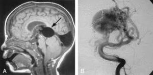
 Vein of Galen aneurysmal malformations (VGAMs) and Vein of Galen aneurysmal dilations (VGADs) are the most frequent arteriovenous malformations in infants and fetuses.
A VGAM consists of a tangled mass of dilated vessels supplied by an enlarged artery.
A fetal vein of Galen malformation is a rare congenital intracranial vascular anomaly characterized by abnormal arteriovenous shunting between cerebral arteries: typically choroidal and subependymal arteries, and the persistent median prosencephalic vein of Markowski, which is the embryologic precursor to the vein of Galen.
This results in a dilated midline venous structure located posterior to the third ventricle, visible on fetal imaging as a large, turbulent vascular sac.
The malformation increases greatly in size with age.
Dilation of the great cerebral vein of Galen is a secondary result of the force of arterial blood either directly from an artery via an arteriovenous fistula or by way of a tributary vein that receives the blood directly from an artery.
There is usually a venous anomaly downstream from the draining vein that, together with the high blood flow into the great cerebral vein of Galen causes its dilation.
The right sided cardiac chambers and pulmonary arteries also develop mild to severe dilation.
Malformations often lead to cardiac failure, cranial bruits, hydrocephaly, and subarachnoid hemorrhage in neonates.
Heart failure is due to the size of the arteriovenous shunt that can steal 80% or more of the cardiac output, with large volumes of blood under high pressure returning to the right heart and pulmonary circulation.
It is also the most common cause of death in such patients.
Associated conditions: rare non-developmental syndromes also directly or indirectly affect the great cerebral vein of Galen, although they are extremely rare: superior vena cava syndrome (SVCS), and thrombosis of the lateral sinus, superior sagittal sinus, internal jugular vein, or of the great cerebral vein of Galen itself.
10%-30% of vein of Galen aneurysmal malformations are associated with deleterious heterozygous mutations of EPHB4.
Testing for a malformed vein of Galen is indicated when a patient has heart failure which has no obvious cause.
Diagnosis is suggested by signs such as cranial bruits and symptoms such as expanded facial veins.
The vein of Galen can be visualized using ultrasound or Doppler, and will be noticeably enlarged.
Ultrasound is a particularly useful tool for vein of Galen malformations because so many cases occur in infancy and ultrasound can make diagnoses prenatally.
Many cases are diagnosed only during autopsy as congestive heart failure occurs very early.
Five patterns of Galenic arteriovenous malformations have been described:
Pattern 1 Many vessels, including anterior cerebral arteries, thalamic perforating arteries, and superior cerebellar arteries discharge into the vein of Galen.
Pattern 2 A single posterior choroidal artery drains into the vein of Galen.
Pattern 3 One or both posterior choroidal and one or both anterior cerebral arteries drain directly into the Galenic system.
Pattern 4 An angiomatous network of posterior choroidal and thalamic perforating arteries enter the Vein of Galen directly.
Pattern 5 A high flow arteriovenous malformation in the right inferior frontal lobe drains via the inferior sagittal sinus and pericallosal vein into the Vein of Galen.
These malformations develop in utero by the persistence of fistulae between primitive pia arachnoidal arteries and pial veins that cross each other at right angles.
The primitive Galenic system and the primitive choroidal system lie close together, an arteriovenous malformation involving the primitive choroidal system will inevitably involve the Galenic vein.
Larger arteriovenous shunts correlate with greater hemodynamic effects and earlier symptom onset.
Small arteriovenous shunts correlate with greater local mass effect causing progressive neurological impairment.
Treatment depends on the anatomy of the malformation as determined by angiography or Magnetic Resonance Imaging (MRI).
The head’s circumference measurements should be obtained regularly and monitored carefully to detect hydrocephalus.
Neurosurgical procedures to relieve hydrocephalus with a ventriculoperitoneal shunt may be required in some infants.
The fistulous arteries feeding into the Vein of Galen must be blocked, thereby reducing the blood flow into the vein.
Open surgery has a high morbidity and mortality making endovascular embolization the preferred method of treatment.
There have been several reported cases of arteriovenous malformations recurring.
Radiotherapy, also called radiosurgery, involves the use of focused beams to damage the blood vessel.
Such radiotherapy is often not pursued as a treatment because the effects of the procedure can take months or years and there is risk of damaging adjacent brain tissue.
Surgery treatment is not always an option when the anatomy of the malformation creates too much of a risk.
Endovascular procedures have made many cases, which were not surgically accessible, treatable.
Endovascular treatments involve delivering drugs, balloons, or coils to the site of the malformation through blood vessels via catheters.
Endovascular treatments work by limiting blood flow through the vein.
Risks of complications from endovascular treatments:the wall of the vein can be damaged during the procedure, and emboli can become dislodged and travel through the vascular system.
Color-flow imaging and pulsed Doppler ultrasonography provides anatomical and pathophysiological information regarding hemodynamics and intracranial blood flow.
These methods provided a reliable, noninvasive means to evaluate the effectiveness of therapy and the need for further treatment in neonates with Vein of Galen malformations.
Seizures usually are managed with antiepileptic medications.
The complications usually associated with vein of Galen malformations are usually intracranial hemorrhages.
Over half the patients with VGAM have a malformation that cannot be corrected.
Patients frequently die in the neonatal period or in early infancy.
77% of untreated cases result in mortality.
Even after surgical treatment, the mortality rate remains as high as 39.4%.
Most cases occur during infancy when the mortality rates are at their highest.
The malformation develops between the 8th and 11th weeks of gestation due to aberrant morphogenesis of the cerebral vasculature, leading to high-flow arteriovenous fistulas.
Prenatal diagnosis is typically made in the third trimester using Doppler ultrasound and fetal MRI, which can delineate the vascular anatomy, assess for secondary brain injury, and evaluate cardiac status.
Common associated clinical findings include: ventriculomegaly, cardiomegaly, tricuspid regurgitation, and signs of high-output cardiac failure, which are important prognostic indicators.
Genetic studies have identified loss-of-function mutations in EPHB4 as a cause in some cases, implicating disrupted vascular development pathways.
Prognosis depends on the degree of cerebral and cardiac compromise; significant morbidity and mortality are associated with severe cardiac failure and brain injury.