Focused ultrasound/histotripsy
High intensity focused ultrasound (HIFU) can be used to destroy tissue in the body.
HIFU beams are precisely focused on a small region of diseased tissue to locally deposit high levels of energy.
An acoustic lens is used to focus sound to a small point in the body.
The sound propagates through many layers of tissue, and only tissue at the focus is destroyed.
High-intensity focused ultrasound (HIFU), is an incisionless therapeutic technique that uses non-ionizing ultrasonic waves to heat or ablate tissue.
It can be used to increase the flow of blood or lymph or to destroy tissue, such as tumors, via thermal and mechanical mechanisms.
Compared to ultrasonic imaging, it uses lower frequencies and continuous, rather than pulsed, to achieve the necessary thermal doses.
However, pulsed waves may also be used if mechanical rather than thermal damage is desired.
Acoustic lenses are often used to achieve the necessary intensity at the target tissue without damaging the surrounding tissue.
HIFU is combined with other imaging techniques such as medical ultrasound or MRI to enable guidance of the treatment and monitoring.
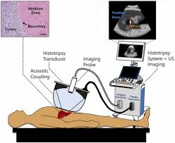
Histotripsy mechanically destroys tissue through cavitation.
HIFU is used to non-invasively heat or ablate tissue deep in the body without the need for an incision, with it main applications being the destruction of tissue caused by hyperthermia, increasing perfusion and physical therapy.
It is being used to treat tumors of the liver, with clinical trials underway for other sites.
The use of ultrasound in the treatment of musculoskeletal conditions is another use in the physiotherapy setting.
A focused ultrasound system can treat essential tremor, neuropathic pain, and Parkinsonian tremor.
HIFU may be effective for treating prostate cancer.
HIFU has been studied in liver cancer with high response rates and positive outcome.
During the treatment of metastasized liver cancer with HIFU, immune responses have been observed in locations that are distant from the focal region.
A clinical trial of the histotripsy form of HIFU for liver tumors, researchers demonstrated a 95% success rate.
Treatment of benign prostatic hyperplasia by HIFU from inside the intestine has turned out to be unsuccessful.
Focused ultrasound may be used to generate highly localized heating to treat cysts and tumors.
Procedures generally use lower frequencies than medical diagnostic ultrasound (from 0.7 to 2 MHz), but higher the frequency means lower the focusing energy. HIFU treatment is often guided by MRI.
Focused ultrasound may be used to dissolve kidney stones by lithotripsy.
Ultrasound may be used for cataract treatment by phacoemulsification.
The focused temperature at tissue will rise to between 65 and 85 °C, destroying the diseased tissue by coagulative necrosis.
If tissue is elevated above the threshold of 60 °C for longer than 1 second this necrotic process is irreversible.
With focused beams, a very small region of heating can be achieved the part of shallow deep in tissues (usually on the order of 2~3 millimeters).
Thermal doses of 120-240 min at 43 °C coagulate cellular protein and leads to irreversible tissue destruction.
At high acoustic intensities, cavitation microbubbles form and interact with the ultrasound field.
Microbubbles produced in the field oscillate and grow and can eventually implode.
Stable cavitation creates microstreaming which induces high shear forces on cells and leads to apoptosis.
The ultrasound beam can be focused in these ways:
Geometrically, for example with a lens or with a spherically curved transducer.
Electronically, by adjusting the relative phases of elements in an array of transducers.
Beam delivery consists of beam steering and image guidance.
The beam has the ability to pass through overlying tissues without harm and focus on a localized area with size limit of 2–3 mm, that is determined the clinical frequency of the ultrasound.
Following ablation a distinct boundary forms between healthy and necrotic tissue with a width less than 50 microns.
Pre-operative imaging with CT and MRI, are usually used to identify general the target anatomy.
Real-time imaging, on the other hand, is necessary for safe and accurate noninvasive targeting and therapy monitoring: MRI and Medical ultrasound.
Magnetic Resonance guided Focused Ultrasound Surgery (MRgFUS) is a 3D imaging technique which features high soft tissue contrast and provides information about temperature, thus allowing to monitor ablation.
Ultrasound is ideal for image guidance is it verifies the acoustic window in real time since it is the same modality as the therapy.
Treatment outcomes can be estimated in real time through visual inspection of hyperechoic changes in standard B-mode images.