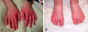
Erythromelalgia is a vascular peripheral pain disorder in which blood vessels, usually in the lower extremities or hands, are episodically blocked and then become hyperemic and inflamed.
 It is associated with severe burning pain in the small fiber sensory nerves and skin redness.
The attacks are periodic and are commonly triggered by heat, pressure, mild activity, exertion, insomnia or stress.
Erythromelalgia may occur either as a primary or secondary disorder.
Secondary erythromelalgia can result from small fiber peripheral neuropathy of any cause: polycythemia vera, essential thrombocythemia, hypercholesterolemia, mushroom or mercury poisoning, and some autoimmune disorders.
Primary erythromelalgia is caused by mutation of the voltage-gated sodium channel α-subunit gene SCN9A.
SCN9A mutations enhance the function of NaV1.7 sodium channels, which are preferentially expressed within peripheral neurons.
NaV1.7 mutants channels, from families with inherited erythromelalgia (IEM), make dorsal root ganglion neurons hyper excitable, demonstrating the mechanistic link between these mutations and pain.
It is established NaV1.7 gain-of-function mutations are the molecular basis for erythromelalgia.
Primary erythromelalgia may be classified as either familial or sporadic, with the familial form inherited in an autosomal dominant manner, and further classified as either juvenile or adult onset.
The genetic cause of the juvenile and sporadic adult onset forms is often known, this is not the case for the adult onset familial form.
An epidemic form of erythromelalgia has been viewed as a different form of non-inherited primary erythromelalgia and affects mainly teenage girls in middle schools.
The epidemic disease is characterized by burning pain in the toes and soles of the feet, accompanied by foot redness, congestion, and edema; a few patients may have fever, palpitations, headache, and joint pain.
The epidemic form may have a common cold before the onset of erythromelalgia.
The most prominent symptoms of erythromelalgia are episodes of erythema, swelling, a painful deep-aching of the soft tissue and tenderness, along with a painful burning sensation primarily in the extremities.
These symptoms are often symmetric.
These symptoms affect the lower extremities more frequently than the upper extremities.
Symptoms may also affect the ears and face.
For secondary erythromelalgia, attacks typically occur before and are precipitated by the underlying primary condition.
For primary erythromelalgia, attacks can last from an hour to months at a time and occur infrequently to frequently with multiple times daily.
Attacks most frequently occur at night, potentially interfering with sleep.
Common triggers for daytime episodes: exertion, heating of the affected extremities, and alcohol or caffeine consumption, and any pressure applied to the limbs.
In some patients sugar and even melon consumption have also been known to provoke attacks.
With primary erythromelalgia wearing shoes or socks can generate heat known to produce erythromelalgia attacks.
The coexistence of erythromelalgia and Raynaud’s phenomenon is rare.
Symptoms may present gradually and incrementally.
Sometimes it takes years to become intense enough for patients to seek medical care, while other cases symptoms emerge full blown with onset.
Epidemic erythromelalgia is characterized by burning pain in the toes and soles of the feet, foot redness, congestion, and edema following a cold or pharyngitis: it may be associated with fever, palpitations, headache, and joint pain.
EM is caused by an underlaying small fiber neuropathy.
Primary erythromelalgia seems to consist of neuropathological and microvascular alterations.
Secondary erythromelalgia cause is poorly understood and may be specific to the underlying primary condition.
Primary erythromelalgia is a better understood autosomal dominant disorder.
The neuropathological process of primary erythromelalgia arise from hyperexcitability of C-fibers in the dorsal root ganglion.
Specifically, nociceptors appear to be the primarily affected neurons in these fibers, with hyperexcitability resulting in the severe burning pain experienced by patients.
The neuropathological symptoms is a result of hyperexcitability, while microvascular alterations in erythromelalgia are due to hypoexcitability.
The sympathetic nervous system controls cutaneous vascular tone and altered response of this system to stimuli such as heat likely results in the observed microvascular symptoms.
These changes in excitability are typically due to mutation of the sodium channel NaV1.7.
Several medications: verapamil and nifedipine, ergot derivatives such as bromocriptine and pergolide, have been associated with medication-induced erythromelalgia.
The consumption of two species of related fungi, has led to several cases of mushroom-induced erythromelalgia.
There are 10 known mutations in the voltage-gated sodium channel α-subunit NaV1.7 encoding gene, SCN9A.
This channel is expressed primarily in nociceptors of the dorsal root ganglion and the sympathetic ganglion neurons.
This results in channels that are open for a longer of period of time, producing larger and more prolonged changes in membrane potential.
Some of these mutant channels have been expressed in dorsal root ganglion (DRG) or sympathetic neurons.
An effective treatment for erythromelalgia symptoms is cooling of the affected area.
Activation of wild-type channels is unaffected by cooling.
Erythromelalgia is a difficult to diagnose as there are no specific tests available.
However, reduced capillary density has been observed microscopically during flaring.
Reduced capillary perfusion is noted.
When a patient elevates their legs a reversal, from red to pale, in skin color occurs.
Quantitative sensory nerve testing, laser evoked potentials, sweat testing and epidermal sensory nerve fiber density tests are objective tests for small fiber sensory neuropathy that are available.
Some diseases present with symptoms similar to erythromelalgia: Complex regional pain syndrome presents with severe burning pain and redness except these symptoms are often unilateral and may be proximal instead of purely or primarily distal.
Erythromelalgia is sometimes caused by other disorders Myeloproliferative disease Hypercholesterolemia Autoimmune disorder Small fiber peripheral neuropathy Fabry’s disease Mercury poisoning Mushroom poisoning Obstructive Sleep Apnea Sciatica Some medications, such as fluoroquinolones, bromocriptine, pergolide, verapamil, and ticlopidine
Treatment: For secondary erythromelalgia, treatment of the underlying primary disorder is the most primary method of treatment.
Aspirin may reduce symptoms of erythromelalgia, but there is rare evidence that this is effective.
Mechanical cooling of the limbs by elevating them can help or managing the ambient environment.
Flares occur due to sympathetic autonomic dysfunction of the capillaries.
The pain that accompanies it is severe.
Patients are strongly advised not to place the affected limbs in cold water to relieve symptoms as it precipitates problems causing damage to the skin and ulceration often intractable due to the damaged skin.
A possible reduction in skin damage may be accomplished by enclosing the flaring limb in a thin, heat transparent, water impermeable, plastic food storage bag.
Primary erythromelalgia management is symptomatic, i.e. treating painful symptoms only.
Specific management tactics include avoidance of attack triggers such as: heat, change in temperature, exercise or over exertion, alcohol and spicy foods.
A cool environment is helpful in keeping the symptoms in control, the use of cold water baths is strongly discouraged.
Patients find relief by cooling the skin. All patients must be notified to not apply ice directly on to the skin, since this can cause maceration of the skin, nonhealing ulcers, infection, necrosis, and even amputation in severe cases.
Clinical studies have demonstrated the efficacy of IV lidocaine or oral mexilitin, misoprostol, gabapentin, venlafaxine and oral magnesium.
A combination of drugs such as duloxetine and pregabalin is an effective way of reducing the stabbing pains and burning sensation symptoms of erythromelalgia in conjunction with the appropriate analgesia.
Antihistamines may give some relief.
Most people with erythromelalgia never go into remission and the symptoms are ever present at some level.
Others get worse, or the EM is eventually a symptom of another disease such as systemic scleroderma.
Ketamine topical creams may help manage pain on a long-term basis.
Erythromelalgia can result in a deterioration in quality of life:inability to function in a work place, lack of mobility, depression, and is socially alienating; much greater education of medical practitioners is needed.
Mild sufferers may find pain relief with tramadol or amitriptyline.
With more severe and widespread EM symptoms, however, may obtain relief only from opioid drugs.
The mean of all the studies combined results in an EM estimation incidence of 4.7/100,000 with a mean of 1 : 3.7 of the male to female ratio, respectively.
This last study has an estimation that is at least ten times higher than the prevalence previously reported.
This study recruited individuals based on self-identification of symptoms (after self-identification, patients were invited for an assessment of an EM diagnosis), instead of participants that are identified through secondary and tertiary referrals as in the other studies.
Epidemic EM appears quite common in female middle school students of southern China, possi ly due to a sharp decline in temperature following by a rapid increase of temperature.
It has been postulated that epidemic erythromelalgia might be related to a poxvirus (ERPV) infection.
Erythromelalgia and erythermalgia on the basis of responsiveness to erythromelalgia (platelet-mediated and aspirin-sensitive), primary erythermalgia, and secondary erythermalgia.
The primary/idiopathic form of erythromelalgia is not associated with any other disease process and can be either early onset in children or adult onset.
Secondary erythromelalgia as being associated with another disease, often related to a myeloproliferative disorder and has also seen cases of: hypertension, diabetes mellitus, rheumatoid arthritis, gout, systemic lupus erythematosus, multiple sclerosis, astrocytoma of the brain, vasculitis, and pernicious anemia.
