
Obstetric ultrasonography, or prenatal ultrasound, is the use of medical ultrasonography in pregnancy.
Sound waves are used to create real-time visual images of the developing embryo or fetus in the uterus.
OU can provide a variety of information about the health of the mother, the timing and progress of the pregnancy, and the health and development of the embryo or fetus.
The International Society of Ultrasound in Obstetrics and Gynecology recommends that pregnant women have routine obstetric ultrasounds between 18 weeks’ and 22 weeks’ gestational age to confirm pregnancy dating, to measure the fetus so that growth abnormalities can be recognized quickly later in pregnancy, and to assess for congenital malformations and multiple pregnancies.
It is recommende that pregnant patients who desire genetic testing have obstetric ultrasounds between 11 weeks’ and 13 weeks 6 days’ gestational age.
Performing an ultrasound at this early stage of pregnancy can more accurately confirm the timing of the pregnancy, and can also assess for multiple fetuses and major congenital abnormalities at an earlier stage.
Obstetric ultrasound before 24 weeks’ gestational age can significantly reduce the risk of failing to recognize multiple gestations and can improve pregnancy dating to reduce the risk of labor induction for post-dates pregnancy.
There is no difference, however, in perinatal death or poor outcomes for infants.
Transvaginal ultrasonography – Ultrasound is performed through the vagina.
Transabdominal ultrasonography – Ultrasound is performed across the abdominal wall or through the abdominal cavity.
In normal state, each body tissue type, such as liver, spleen or kidney, has a unique echogenicity.
Fortunately, gestational sac, yolk sac and embryo are surrounded by hyperechoic or brighter body tissues.
Traditional obstetric sonograms are done by placing a transducer on the abdomen of the pregnant woman.
Transvaginal sonography, is done with a probe placed in the woman’s vagina.
Transvaginal scans usually provide clearer pictures during early pregnancy and in obese women.
Doppler sonography which detects the heartbeat of the fetus.
Doppler sonography can be used to evaluate the pulsations in the fetal heart and bloods vessels for signs of abnormalities.
There are no definitive studies linking ultrasound to any adverse medical effects.
A gestational sac can be reliably seen on transvaginal ultrasound by 5 weeks’ gestational age (approximately 3 weeks after ovulation).
The embryo can be seen by the time the gestational sac measures 25 mm, about five and a half weeks.
The heartbeat is usually seen on transvaginal ultrasound by the time the embryo measures 5 mm, but may not be visible until the embryo reaches 19 mm, around 7 weeks’ gestational age.
Most miscarriages also happen by 7 weeks’ gestation.
The rate of miscarriage, especially threatened miscarriage, drops significantly after normal heartbeat is detected, and after 13 weeks.
Contents in the cavity of the uterus seen at approximately 5 weeks of gestational age
In the first trimester, a standard ultrasound examination typically includes:
Gestational sac size, location, and number
Identification of the embryo and/or yolk sac
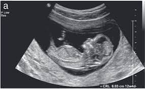
Measurement of fetal length (known as the crown-rump length)
Fetal number, including number of amnionic sacs and chorionic sacs for multiple gestations
Embryonic/fetal cardiac activity
Assessment of embryonic/fetal anatomy appropriate for the first trimester
Evaluation of the maternal uterus, tubes, ovaries, and surrounding structures
Evaluation of the fetal nuchal fold, with consideration of fetal nuchal translucency assessment
In the second trimester, a standard ultrasound exam typically includes:
Fetal number, including number of amnionic sacs and chorionic sacs for multiple gestations
Fetal cardiac activity
Fetal position relative to the uterus and cervix
Location and appearance of the placenta, including site of umbilical cord insertion when possible
Amniotic fluid volume
Gestational age assessment
Fetal weight estimation
Fetal anatomical survey
Evaluation of the maternal uterus, tubes, ovaries, and surrounding structures when appropriate
Dating and growth monitoring
Biparietal diameter is taken as the maximal transverse diameter of in a visualization of the horizontal plane of the head.
Establishing gestational age is critical for guiding obstetrical care, including the timing of prenatal visits, laboratory testing, administration of medications, vaccinations, and delivery time as well as neonatal care, including care for newborn resuscitation and neonatal intensive care.
Obstetrical ultrasound is the criterion standard tool for establishing pregnancy, viability and dating, and for assessing fetal anatomy, growth, and well-being.
Gestational age is usually determined by the date of the woman’s last menstrual period, and assuming ovulation occurred on day fourteen of the menstrual cycle.
Ultrasound scans offer an alternative method of estimating gestational age.
The most accurate measurement for dating is the crown-rump length of the fetus, which can be done between 7 and 13 weeks of gestation.
After 13 weeks of gestation, the fetal age may be estimated using the biparietal diameter, the head circumference, the length of the femur, the crown-heel length and other fetal parameters.
Dating is more accurate when done earlier in the pregnancy.
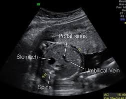
The abdominal circumference of the fetus may also be measured, giving an estimate of the weight and size of the fetus and is important when doing serial ultrasounds to monitor fetal growth.
The sex of the fetus may be discerned by ultrasound as early as 11 weeks’ gestation.
After 13 weeks’ gestation, there is a high accuracy of between 99% and 100% is possible if the fetus does not display intersex external characteristics.
The accuracy of fetal sex discernment depends on:
Gestational age Precision of sonographic machine Expertise of the operator Fetal posture Ultrasonography of the cervix
Obstetric sonography is useful in the assessment of the cervix in women at risk for premature birth.
A short cervix preterm is associated with a higher risk for premature delivery.
At 24 weeks’ gestation, a cervix length of less than 25 mm defines a risk group for spontaneous preterm birth:the shorter the cervix, the greater the risk.
Cervical measurement on ultrasound is helpful in patients with preterm contractions, as those whose cervical length exceeds 30 mm are unlikely to deliver within the next week.
Routine pregnancy sonographic scans are performed to detect developmental defects before birth: checking the status of the limbs and vital organs.
The most common US test uses a measurement of the nuchal translucency thickness.
Although 91% of fetuses affected by Down syndrome exhibit this defect, 5% of fetuses flagged by the test do not have Down syndrome.
Ultrasound may also detect fetal organ anomaly.
Usually scans for this type of detection are done around 18 to 23 weeks of gestational age and is called the anatomy scan.
Such scans are beneficial because ultrasound enables assessing the developing fetus in terms of morphology, bone shape, skeletal features, fetal heart function, volume evaluation, fetal lung maturity, and general fetus well being.
Second-trimester ultrasound screening for aneuploidies is looking for soft markers and some predefined structural abnormalities.
Current evidence indicates that diagnostic ultrasound is safe for the unborn child.
In a randomized trial, the children with greater exposure to ultrasound had a reduction in perinatal mortality, and was attributed to the increased detection of anomalies in the ultrasound group.
Randomized controlled trials have reported no association between Doppler exposure and birth weight, Apgar scores, and perinatal mortality.
