
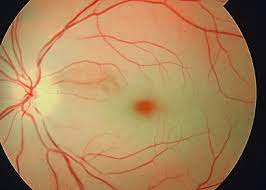
 Central retinal artery occlusion (CRAO) is an ophthalmic emergency because irreversible ischemic damage to the retina can occur in as little as 90 minutes.
It is considered an ocular stroke and the risk of ischemic stroke with large vessel involvement is particularly increased in the first one to four weeks after a diagnosis of a CRAO.
Patients may present with sudden, painless decreased vision in one eye over a matter of seconds,with an afferent pupillary defect.
It may be partial vision loss if one of the smaller distal branches are affected.
Patients may have experienced intermittent transient, painless vision loss.
Risk factors for CRAO include cigarette smoking, hypertension, high body mass index, high serum lipid levels, diabetes, cardiac disease, hypercoagulable conditions such as antiphospholipid antibody syndrome and autoimmune conditions.
Clinically blanched, non perfused retina, and a cherry red spot due to bright red appearance of the fovea on fundoscopic examination may be apparent.
Fundoscopy usually show signs of acute ischemia, including retinal pallor a cherry, red spot, segmented blood flow, and attenuated, retinal arteries, although the fundus may appear normal.
In the acute phase optical coherence tomography usually shows inner retinal, hyperreflexivity and thickening.
Central retinal artery occlusion is typically caused by embolism from a carotid plaque or a cardiac thrombus, but can result from any type of ischemic event.
A boxcar pattern can be seen in the retinal arteries and veins due to segmented blood flow.
No proven therapy exists for retinal artery occlusion.
Digital massage to dislodge the clot to a distal branch and reduce ischemia, anterior chamber paracentesis, and carbogen inhalation have been suggested, all with poor visual outcomes.
Therapy with full dose aspirin and evaluation with carotid imaging and neurological workup, MRI of the brain, echocardiography, Holter monitoring should be performed.
Giant cell arthritis should be ruled out.
Patients should have diet alterations, smoking cessation, and initiation of statin therapy in the case of dyslipidemia.
Intravenous tenecteplase administered within 4.5 hours after onset of central retinal artery occlusion does not result in significantly greater vision recovery at 30 days than oral aspirin, but is associated with the significant serious safety concerns: unlike the management of acute ischemic stroke findings do not suggest the routine use of thrombolytic therapy to treat central retinal artery occlusion.