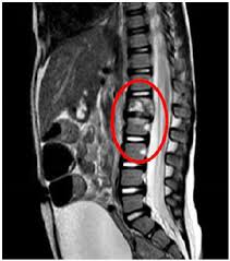
Pott disease is tuberculosis of the spine.
It is usually the result of haematogenous spread from other sites, often the lungs.
The lower thoracic and upper lumbar vertebrae areas of the spine are most often affected.
It manifests as a tuberculous arthritis of the intervertebral joints.
The infection can spread from two adjacent vertebrae into the adjoining intervertebral disc space.
If only one vertebra is affected, the disc is normal.
If two are involved, the disc, which is avascular, cannot receive nutrients, and collapses.
Caseous necrosis of the disc leads to vertebral narrowing and eventually to vertebral collapse and spinal damage.
A dry soft-tissue mass often forms and superinfection is rare.
Spread from the lumbar vertebrae to the psoas muscle can cause causing abscesses, but uncommon.
Diagnostic tests.
Complete blood count: leukocytosis
Elevated erythrocyte sedimentation rate: >100 mm/h
Tuberculin skin test- positive in 84–95% of patients with Pott disease who are not infected with HIV.
Radiographic changes associated with Pott disease present relatively late findings.
Radiographic changes are characteristic of spinal tuberculosis on plain radiography:
Lytic destruction of anterior portion of vertebral body
Increased anterior wedging
Collapse of vertebral body
Reactive sclerosis on a progressive lytic process
Enlarged psoas shadow with or without calcification.
Vertebral end plates are osteoporotic.
Intervertebral disks may be shrunken or destroyed.
Vertebral bodies show variable degrees of destruction.
Fusiform paravertebral shadows suggest abscess formation.
Bone lesions may occur at more than one level.
MANAGEMENT:
Nonoperative – antituberculous drugs
Immobilization of the spine: braces and collars.
Surgery may be necessary, especially to drain spinal abscesses or debride bony lesions fully or to stabilize the spine.
Surgery should not be recommended routinely and clinicians have to selectively judge and decide on which patients to operate.
